 |
| November 14, 2017 | Volume 13 Issue 42 |
Designfax weekly eMagazine

Archives
Partners
Manufacturing Center
Product Spotlight
Modern Applications News
Metalworking Ideas For
Today's Job Shops
Tooling and Production
Strategies for large
metalworking plants
Great Engineering: $10 microchip turns 2D ultrasound machine into 3D imaging device
Technology that keeps track of how your smartphone is oriented can now give $50,000 ultrasound machines many of the 3D-imaging abilities of their $250,000 counterparts -- for the cost of a $10 microchip.
In a presentation at the American College of Emergency Physicians Research Forum on Oct. 31, doctors and engineers from Duke University and Stanford University showed how their snap-on invention can make 2D ultrasound machines much more powerful. With a few cents' worth of 3D-printed materials, a microchip common to smartphones, a few wires, and a laptop, a 2D ultrasound machine can capture 3D images of infants' brains with similar quality to MRI or CT scans while the baby is held in a parent's arms.
"With 2D technology you see a visual slice' of an organ, but without any context you may mistake it for another part or mistake one disease process or injury for another," said Joshua Broder, M.D., emergency physician and director of the emergency medicine residency program at the Duke University School of Medicine. "These are all problems that can be solved with the added orientation and holistic context of 3D technology. Gaining that ability at an incredibly low cost by taking existing machines and upgrading them seemed like the best solution to us."
The idea came to Broder while playing a game of Nintendo Wii with his son. With the game console's ability to accurately track the exact position of the controller, Broder thought to himself, "Why not just duct tape the controller to an ultrasound probe?"
That is the basic idea behind the new device. When physicians use a 2D ultrasound machine, they move and tilt the probe in various directions until they get the view they want. But in the process, they are essentially throwing away perfectly good data. The new device keeps track of the exact orientation of the probe while imaging a patient, so that those images can be stitched together in three dimensions.
"If each image were a playing card, then it would be like putting them all together in order into a deck so you can flip through it," said Broder. "Each image is a slice that makes up the whole."
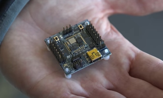
The $10 chip that transforms the basic ultrasound into a 3D imager.
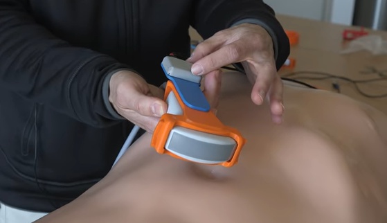
The prototype includes a 3D-printed harness that snaps onto the ultrasound probe to securely attach the position-tracking microchip. That harness then snaps into one of several platforms that restricts its movement to a single axis of rotation.
For example, one platform only allows the probe to swing in an arc, like a door on a hinge. Another only allows it to twist in a circle along its vertical axis. These platforms are meant to allow the orientation of the probe to change while keeping the device centered over one location on a patient.
The microchip is connected to a computer via a USB cord, while the ultrasound device itself is connected through any type of visual cord, be it USB, HDMI, or DVI. As long as the ultrasound machine has a video output -- which nearly all of them do -- the new device can upgrade it to three dimensions.
"We've tried it on all types of ultrasound machines made by GE, Siemens, Philips, SonoSite -- you name it -- and it's worked every time without any previous testing," said Broder. "After a quick arc or twist of the probe, the software churns out a 3D image in a matter of seconds."
To make the sensor-equipped three-dimensional ultrasound probe requires "a few cents worth of plastic and a $10 chip," says Dr. Joshua Broder, the device's inventor. The chip is housed inside the gray plastic add-on.
The invention is currently being tested in clinical trials at both Duke and Stanford, and the study presented at the research forum is a perfect example of its utility.
Some newborns need brain imaging to diagnose congenital problems, but infants are notoriously difficult to scan. MRI machines require stillness over an extended period of time to achieve high resolution, which means either sedating the baby or settling for a lower resolution. CT scans provide excellent 3D images, but they expose the infant to radiation.
Both require the baby be put into a large machine and cost a lot of money.
In the study, physicians were able to take a normal ultrasound probe upgraded with the 3D conversion kit, center it above the infant's head, twist the probe 180 degrees, and get 3D images of similar quality to either CT or MRI. The whole process took a few seconds and was completed while the babies were still in their mothers' arms. "Ultrasound is such a beautiful technology because it's inexpensive, it's portable, and it's completely safe in every patient," said Broder. "And it's brought to the bedside, and it doesn't interfere with patient care."
According to Broder, the 3D upgrade kit is "months away from being ready for prime time." It has come a long way since he envisioned it in 2014, and he attributes its success to the resources available at both Duke and Stanford.
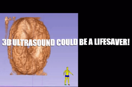
The 3D ultrasound upgrade works so well, you can just make out the name of this Life Savers candy on its gummy surface.
After spending his spare time on it as a labor of love for a year, Broder sought help from Bob Barnes, an instructor in Duke's Masters of Engineering Management program who leads a course on creating biomedical devices. Barnes introduced Broder to the Duke engineers who would come to form the team behind the success of the new device: Matt Morgan, an undergraduate at the time who is now in graduate school at Duke, and Carl Herickhoff and Jeremy Dahl, who were both biomedical engineering instructors and professors at Duke.
Herickhoff and Dahl have since taken positions at the Stanford University School of Medicine, where they continue to develop the device. Their effort is supported by Stanford's branch of the Wallace H. Coulter Foundation, which supports collaborative translational research projects at several universities, including Duke.
The team also had initial help with the patenting and company formation through the Duke Office of Licensing and Ventures. More recent support has come from grants from the Emergency Medicine Foundation and General Electric, which are funding clinical trials for the device's use to locate and identify bleeding and hemorrhages in patients with traumatic shock.
The team believes their current clinical trials and support will allow them to bring their idea to market in only a couple of years. They envision a wide range of benefits, such as bringing 3D imaging to limited-resource areas both at home and around the world to lower the cost of care. They are also working to bridge some of the gaps between their work-around 3D technology and current 3D ultrasound devices, such as the ability to capture a beating heart in motion.
With the resources available to them, Broder has high expectations for the coming years.
"I have been working at Duke since 2005, but my focus had always been on taking care of patients, conducting clinical research with existing imaging technologies, and educating residents," said Broder. "I look back and can't believe that I was as blind as I was to the opportunities of having a world-class university with an engineering school focused on innovation and collaboration right down the street. It really broadened my horizons to what was possible."
Source: Duke University
Published November 2017
Rate this article
View our terms of use and privacy policy